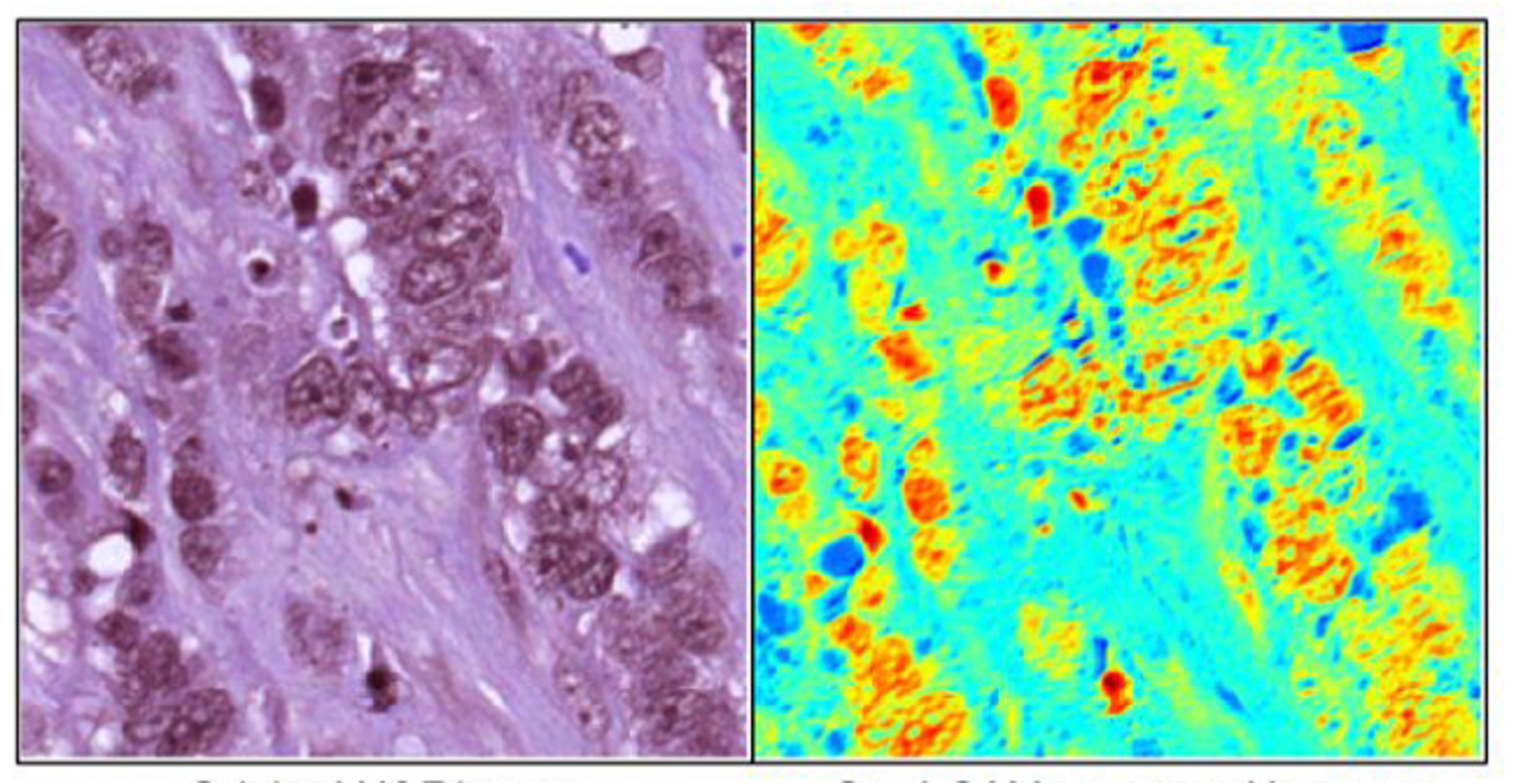
Grad-CAM visualization on tumor regions (Original Hematoxylin & Eosin Image: left, Grad-CAM genreated heatmap: right)
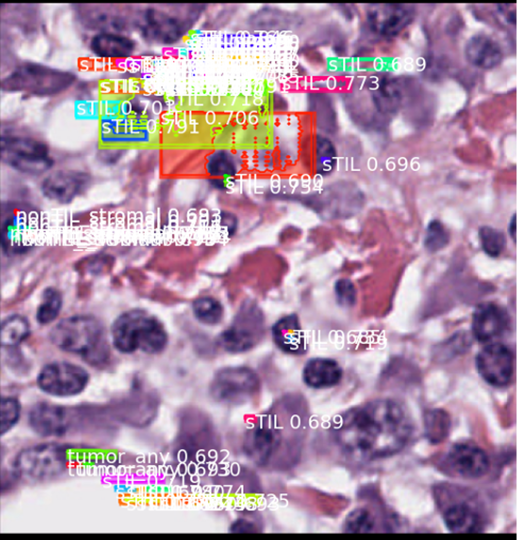
Results of single cell image detection uisng MaskRCNN
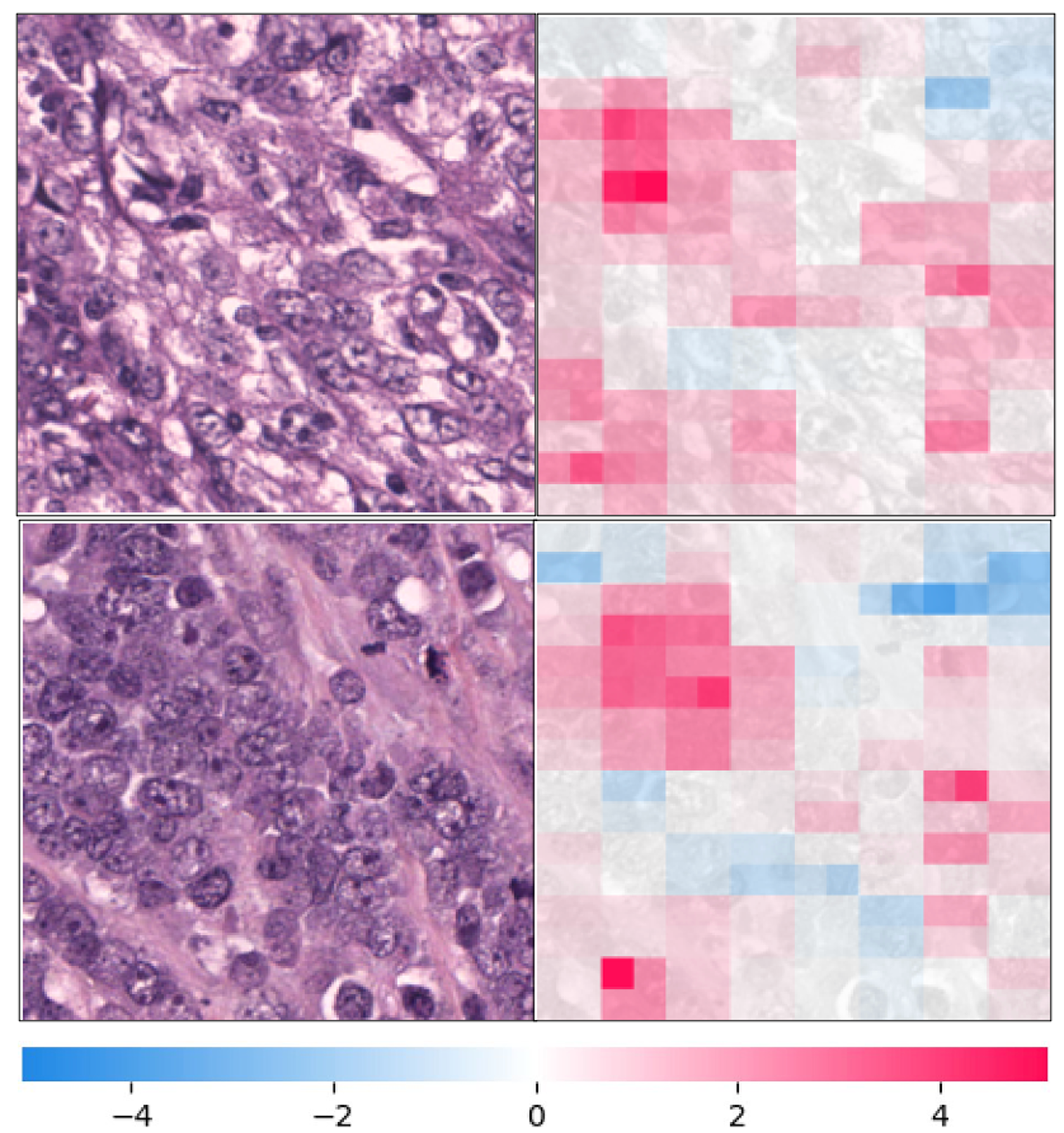
Our project aims to explore human tissue cells digitized by whole slide scanners for a better understanding of complex tumor microenvironments in breast cancer histopathology images, using various deep neural network models. First, we experimented with 70% percentages of tumor cells on image classification using ResNet50, VGG16, and Inception-ResNet. Second, we performed instance image segmentation using Mask-RCNN. Third, we applied two well-known explainable artificial intelligence (AI) techniques including Gradient-weighted Class Activation Mapping (Grad-CAM) and Shapley Additive Explanations (SHAP) to determine the effectiveness of the models.
@inproceedings{jackson2024comprehensive,
          title={Comprehensive Experiments on Breast Cancer Hematoxylin and Eosin-stained Images using UNet},
          author={Jackson, Emily and Le, Faye and Lisbon, Je’Dae and Coleman, Max and Burman, Jordyn and Wonderley, Astrid and Eshaghian, Sepehr and Lee, Sanghoon},
          booktitle={ACMSE 2024},
          year={2024}
        }